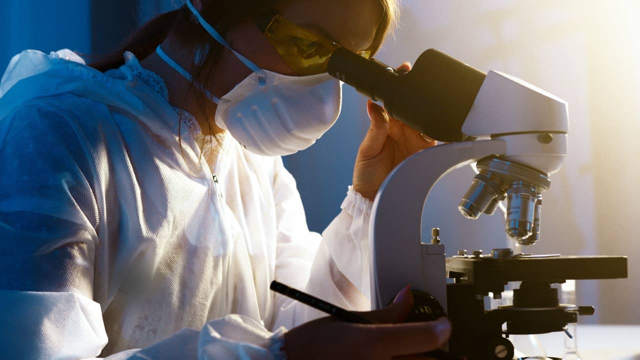
A microscope is lab equipment used to observe tiny objects that can’t be seen through the naked eye. This scientific instrument has lenses that magnify objects such as microorganisms, viruses, or cells depending on their focal strengths. Microscopes come in different types and have one or more lenses with magnification powers of up to 10,000,000X.
Naturally, to hold and support it, a microscope has structural parts and optical sections for enlarging and visualizing the magnified specimen. Most students often struggle to label microscope since to do so, you have to discern the structure of a microscope and its functions. This article will help you understand the microscope parts in detail, as well as why the instrument is important.

Usually, a microscope consists of parts for holding the instrument and optical components for magnification and viewing of the object on the slide. Dive in to find out the 18 parts that make up a microscope.
Structural Parts Of Microscope
Head: It consists of the optical parts in the upper part of the microscope.
Base: It carries the microscopic illuminators and supports them.
Arm: This part connects the tube and head of the microscope to the base, supports the head, and is held when ferrying the microscope.
Optical Parts
Ocular/Eyepiece lens: These are the top lenses used to look through the microscope.
Objective Lenses: These are the main lenses used for object viewing and some face forward, while others are rare. A microscope has around 3 to 4 objective lenses and each lens has a magnification power of 40x to 100x.
Tube: It holds the eyepiece and connects it to the objective lens.
Revolving Nosepiece: Also called a revolving turret. Holds various objective lenses of different magnification powers. Since it can revolve, you can easily rotate it to change the power magnification as desired.
Adjustment Knobs: A coarse adjustment knob is used to focus on the microscope. Found on the microscope arm, the knob moves the stage up and down to bring the object into focus. The fine adjustment knob on the other hand helps get the specimen under a sharp focus. It is located inside the coarse adjustment knob. Both knobs have to be used under low power.
Illuminator: This part of a microscope serves as the source of light when the specimen is on the slide. Located at the base, the illuminator captures light from external sources such as lamplights or sunlight at a low voltage of around 100V. Most of today’s microscopes have in-built lamps.
Stage: A stage is a platform that holds the specimen on the slide for observation. Found halfway up the arm, the stage clips hold the slides by either mechanical or simple slide clips. The mechanical stage allows you to move the slides effortlessly rather than doing it manually through stage control knobs.
Aperture: This is the hole on the microscope stage. Through the aperture, the transmitted light from the source reaches the stage.
Diaphragm/Iris: The diaphragm or Iris is a rotating disk under the stage present in most microscopes. It has holes of different sizes through which it controls the amount of light getting onto the object. It is positioned below the microscope stage and above the condenser lens. Some microscopes have an Abbe condenser that together controls the focus and intensity of the light.
Condenser: Condenser lenses are found under the stage jointly with the diaphragm. These lenses collect and focus the light from the illuminator onto the specimen. They also help in the production of sharp and clear images with a high magnification power of over 400X.
Abbe condensers are found in high-quality microscopes and have the highest magnification of up to 1000X. Condenser Focus Knob moves the condenser up or down to control the light focus on the specimen.
Rack Stop: Rack stop controls how far the stages go and prevents the objective lens from getting too close to the object slide. When it does, it may destroy the specimen. It also prevents the object slide from going too far up and hitting the objective lens.
Also Read – 3 Tips to Help Children Understand What They Read
Uses and Importance of a Microscope
Generally, a microscope is a crucial part of our society that all students have to recognize. Understanding its various parts and functionalities will not just help you achieve high points in the classroom but assist you in the outside world as well. When we see very tiny details that would otherwise have affected people negatively, it helps come with ways of controlling the specimen.
Role of Microscopes in Education
As mentioned earlier, a microscope is highly essential to students in a classroom. Professors use the instrument to teach learners about the structure of tiny objects they can’t see with their naked eyes. Knowing these key details helps students know about different things and their various functions. Such things include animal and plant cells, fungi, and bacteria. Again, microscopes are important in learning about the physics behind mirrors, lenses, and properties of light.

Role of Microscopes in Medicine
If you are looking at a future career in medicine, then this is the best time to start honing your skills. A microscope helps to ascertain the smallest microscopic organisms that can cause diseases. The identification of the microbe assists doctors in prescribing the appropriate antibiotic for the infection. Scientists also inspect genetic makeup like chromosomes for any defects or genetic engineering.
Before Louis Pasteur reported the presence of microscopic organisms, he viewed them under a microscope in the 1860s. Since then, microscopes have continued to help in the diagnosis and treatment of diseases, as well as the prevention and development of vaccines and cures for infections and diseases.
Role of Microscopes in Crime Investigations
With high-quality microscopes in the world of today, anything is possible. Scientists in forensic laboratories use microscopes in their work to study and understand how criminals carry out their criminal activities. In doing so, they place specimens like fibers, blood, or any kind of traces left under examinations.
Scientists who deal with environmental components such as soil or water also use the instrument. Scientists in the engineering field are not left behind since they examine structure components like buildings, dams, or even bridges.


